Shoulder Arthroscopy
- Home
- / Shoulder Arthroscopy
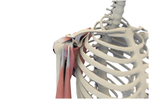
Shoulder Arthroscopy
Dr Rohan Bansal performs the least aggressive and most advanced techniques in arthroscopic and open shoulder surgery. Minimally invasive techniques and use of plasma membranes Rich in Factors (MPRGF) as a healing accelerator
INDICATIONS SHOULDER ARTHROSCOPY:
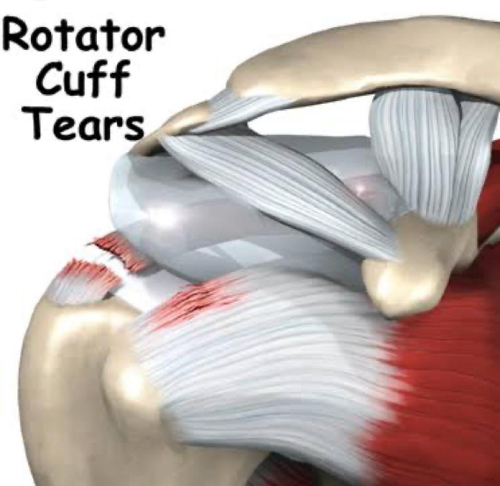
Supraspinatus tears
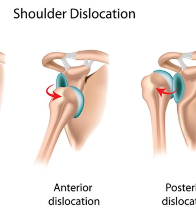
Shoulder Dislocation
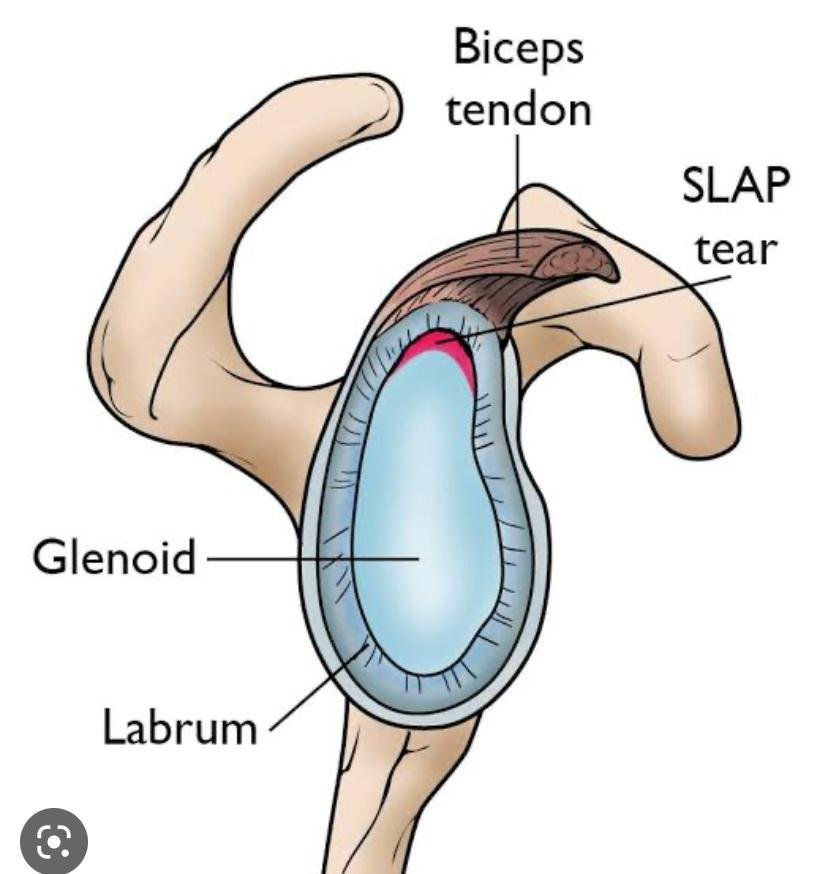
Slap tear
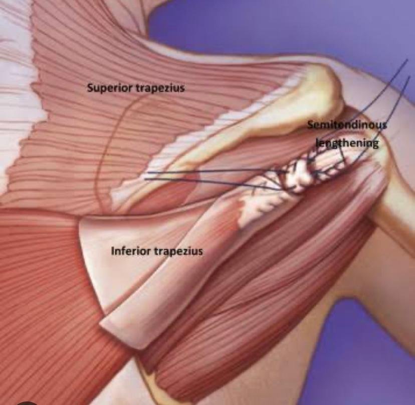
Tendon transfers
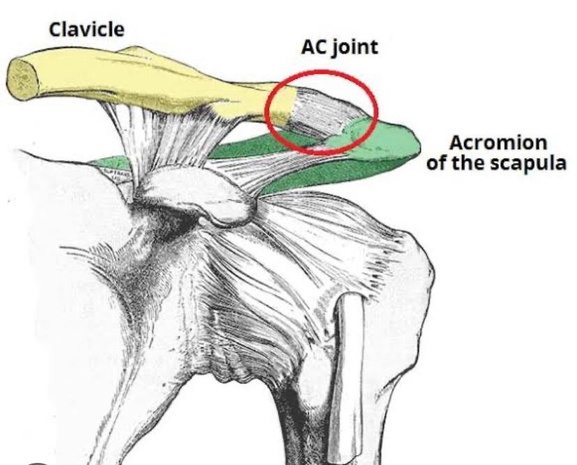
Acromioclavicular joint problems
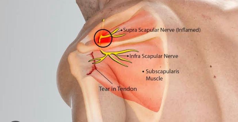
Suprascapular nerve entrapment
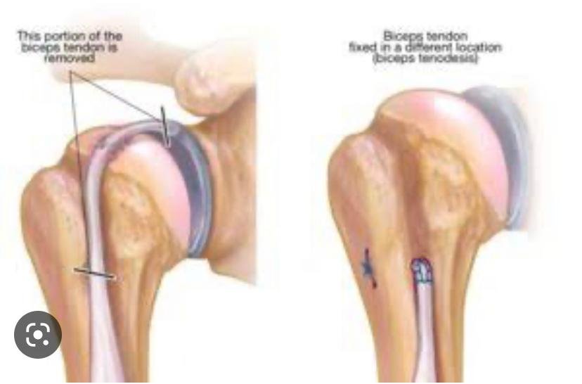
Biceps disorders
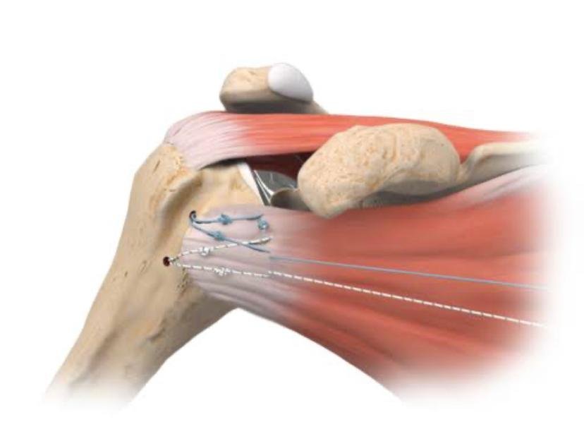
Subscapularis tears
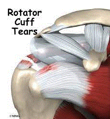
Rotator Cuff Tears
One of the most common conditions in middle-aged people is shoulder pain. A common cause of this pain is a tear or rupture of the rotator cuff. A rotator cuff tear will weaken the shoulder. This means that many of the daily activities such as combing or dressing can be painful and difficult to perform.
WHAT IS THE ROTATOR CUFF?
The rotator cuff helps lift the arm, rotate it, and extend it overhead.
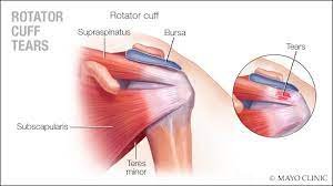
It is made up of muscles and tendons of the shoulder. These muscles cover the head of the upper arm bone (humerus). This “cuff” holds the upper arm bone in the shoulder socket. There are different sizes and shapes of rotator cuff tears. They usually occur in the tendon.
Partial tears. Many tears do not cut through the soft tissue completely.
Full thickness tears. A complete tear will divide the soft tissue into two parts.
HOW ARE TEARS DIAGNOSED?
Your doctor will give you a diagnosis based on your symptoms and physical examination.
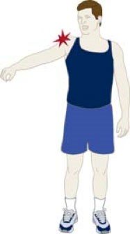
A rotator cuff injury can cause you a lot of pain when you raise your arm to the side.
During the exam, the doctor will move your shoulder in different directions by measuring your range of motion. The doctor also gets important information about the cause of your pain by looking at how well you can move your arm without help. The degree of pain and weakness that a tear causes varies from person to person. Some don’t even realize they have a small rotator cuff tear. Other tests that may help your doctor confirm the diagnosis include:
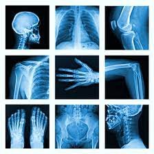
X- rays: Although X-rays do not show rotator cuff tears, they can show other shoulder joint problems. X-rays are usually the first imaging test to be done.
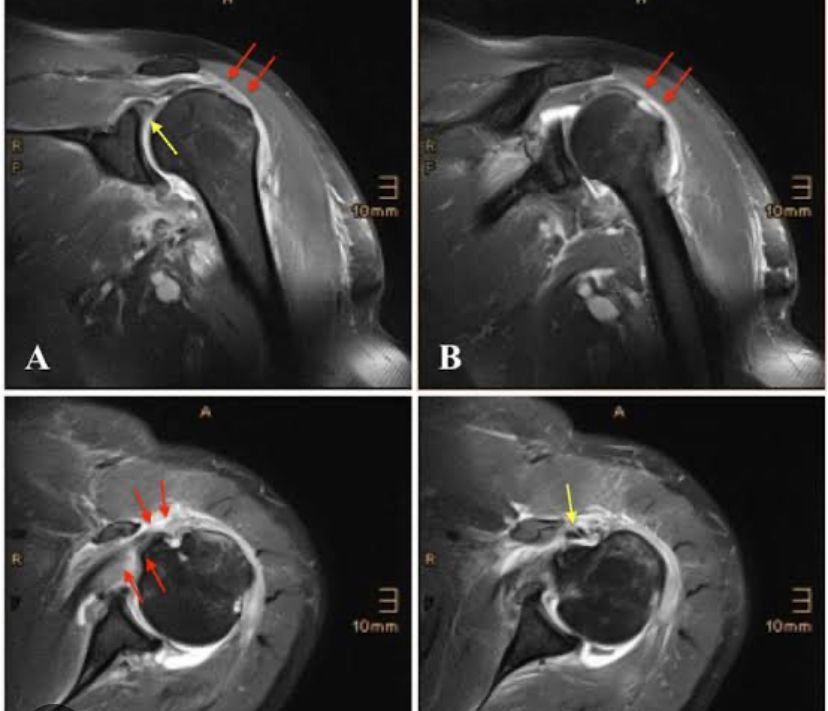
Magnetic resonance imaging (MRI) test and ultrasound. These studies can provide better images of soft tissues such as the rotator cuff tendon. The MRI will be able to show how big the tear is.
WHAT CAUSES ROTATOR CUFF TEARS?
There are two main causes of rotator cuff tears: injury and overuse.
Injury: If you fall on your outstretched arm or lift something very heavy with a sudden movement, you could tear your rotator cuff. This type of tear can occur with other shoulder injuries such as a clavicle fracture or shoulder dislocation.
Overuse: However, most tears are the result of wear and tear on the tendon that occurs slowly over time. This occurs naturally with aging. It can be made worse by overuse, that is, by repeating the same shoulder movements over and over again.
This explains why rotator cuff tears are more common in people over the age of 40 who perform activities that involve repeated movements of the arms above head level. Baseball, tennis, and weightlifting are some examples of sports. Also many jobs and household chores can cause overuse tears. Rotator cuff tears in young people are usually caused by accidents, such as a fall, although overuse tears from sports or work that involve repeated movements of the arms above head level also occur.
How are tears treated?
The goal of treatment is to reduce pain and restore function. When planning treatment, your doctor will take into account your age, activity level, general health, and type of tear.
NON-SURGICAL TREATMENT.
Approximately 50% of patients achieve relief of their symptoms without the need for surgery. Your doctor will be able to start treatment with non-surgical options.
Rest: The first step to recovery is to avoid excessive activity that involves repeated movements of the arms at a height greater than the height of the head.
Non-steroidal and anti-inflammatory drugs: Drugs such as Aspirin and Ibuprofen reduce pain and swelling.
Steroid injections: Cortisone is a very effective anti-inflammatory medication.
Physical therapy: Specific exercises will strengthen your shoulder and restore movement.
REHABILITATION
Whether your treatment involves surgery or not, rehabilitation plays a vital role in getting you back to doing your daily activities. A physical therapy program will help you strengthen and move your shoulder again. Please note that a full recovery takes several months. Although it is a slow process, your commitment to therapy is the most important factor in getting back to doing all the activities you enjoy.
SURGICAL TREATMENT
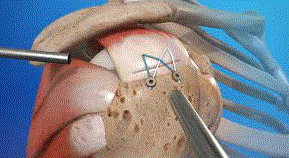
For many people, surgery is the best treatment option. If you are very active and use your arms for sports or work that involves repeated movements of your arms above your head, your doctor will likely consider surgery.
Surgery may be the right option for you for other reasons.
Duration of symptoms:If your symptoms have lasted longer than 6 months, surgery may be suggested.
Major tear : Tears that are larger than 3 centimeters are usually fixed with are usually fixed with surgery.
Weakness: If your shoulder weakness prevents you from doing daily activities, surgery may be the best option.
Trauma: If your tear was caused by a fall or other type of accident, you may have additional injuries. Surgery may be the most effective way to treat all of these injuries.
The type of surgery you need will depend on the size and location of the tear.
SHOULDER instability
Surgical technique for arthroscopic repair involves using All suture and Bioabsorbable anchors in a case of recurrent shoulder dislocation
SHOULDER DISLOCATION
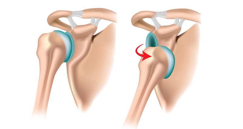
A dislocation is an injury to a joint in which the ball comes out of the socket, similar to a golf ball coming off the golf tee. The shoulder is a “ball-and-socket” joint where the “ball” is the rounded top of the arm bone (humerus) and the “socket” is the cup (glenoid) of the shoulder blade. A layer of cartilage called the labrum cushions and deepens the socket. A shoulder dislocation occurs when the humerus pops out of its socket, either partially or completely. As the body’s most mobile joint, able to move in many directions, the shoulder is most vulnerable to dislocation. A shoulder dislocation may be caused by a sports injury, trauma from a motor vehicle accident, or a fall.
SYMPTOMS OF SHOULDER DISLOCATION
Dislocation causes pain and unsteadiness in the shoulder. The shoulder may be visibly deformed or look out of normal placement. Other symptoms of a dislocated shoulder may include:
Swelling
The muscles in the shoulder may spasm and cause tingling sensations in the neck and down the arm. Complications of a shoulder dislocation may also include muscle tears, tendon or ligament injuries, and blood vessel or nerve damage.
DIAGNOSIS OF SHOULDER DISLOCATION
A shoulder dislocation is diagnosed through a physical examination and a review of symptoms. Additional diagnostic tests may include:
X-ray
MRI scan
TREATMENT OF SHOULDER DISLOCATION
In most cases, the dislocated shoulder can be manipulated back into place by a doctor in a process known as closed reduction. When the shoulder bone is back in place, severe pain normally subsides. The arm and shoulder are then immobilized in a special splint or sling for several weeks as the shoulder heals. Medication may also be prescribed for pain. A shoulder that is severely dislocated or in cases where surrounding ligaments or nerves have been damaged, surgery may be necessary to tighten stretched ligaments or reattach torn ones. After treatment for a shoulder dislocation, when pain and swelling have subsided, physical therapy is recommended to restore the range of motion of the shoulder, strengthen the muscles, and prevent future dislocations. After treatment and recovery, a previously dislocated shoulder may remain more susceptible to reinjury, potentially resulting in chronic shoulder instability and weakness.
ARTHROSCOPIC BANKART REPAIR
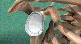
Surgery to repair a Bankart lesion is often performed through arthroscopy. Arthroscopy is a minimally-invasive technique that uses tiny incisions to insert a probe-like camera, allowing the surgeon to fully examine the area before performing corrections.
After making the incisions, the surgeon also inserts specialized instruments through the arthroscope to repair the damage to the shoulder at the exact location of the injury. Any tears in the muscle, tendon, or cartilage will be fixed and any damaged tissue is removed. After the procedure, the incisions are stitched closed.
RECOVERY FROM ARTHROSCOPIC BANKART REPAIR
After arthroscopic Bankart repair, patients will generally be required to keep their arm immobilized in a sling for approximately one month. However, physical therapy will begin on or about day 5 following surgery. In addition, patients will undergo physical therapy for about four months to strengthen the muscle tissue and improve the range of motion in the shoulder. Patients are often restricted from participation in contact sports for a six-month period after surgery, to allow the shoulder to fully heal.
